無料ダウンロード heart anatomy diagram 993320-Heart anatomy diagram unlabeled
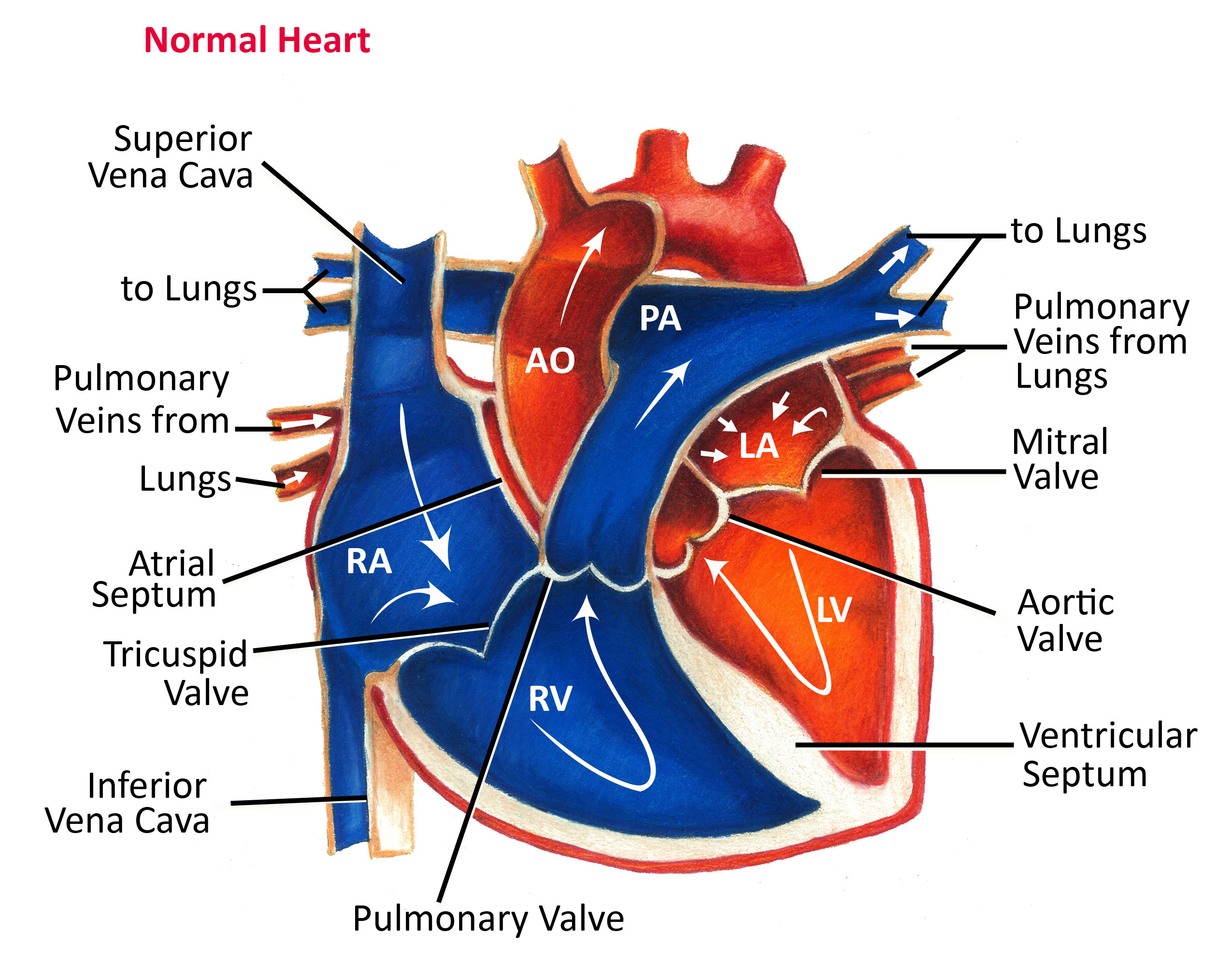
Get the best of Sporcle when you Go OrangeThis adfree experience offers more features, more stats, and more fun while also helping to support Sporcle Thank you for becoming a memberHeart Anatomy and Physiology Heart Nerves Focus topic Heart Anatomy and Physiology The sympathetic and parasympathetic nervous systems that make up the autonomic nervous system are part of the heart's physiology This system does not cause the initiation of the electrical impulses within the cardiac tissue, but, can strongly impact the overall functionDiagram of the blood flow path in the heart Now that blood is freshly oxygenated from its trip to the lungs, it returns to the left atrium of the heart and passively flows through the bicuspid (or

Heart Anatomy Yourheartvalve
Heart anatomy diagram unlabeled
Heart anatomy diagram unlabeled-Because the heart points to the left, about 2/3 of the heart's mass is found on the left side of the body and the other 1/3 is on the right Anatomy of the Heart Pericardium The heart sits within a fluidfilled cavity called the pericardial cavity The walls and lining of the pericardial cavity are a special membrane known as the pericardiumIt consists of the heart, which is a muscular pumping device, and a closed system of vessels called arteries, veins, and capillaries As the name implies, blood contained in the circulatory system is pumped by the heart around a closed circle or circuit of vessels as it passes again and again through the various circulations of the body (on p


Human Heart Anatomy Vector Illustration Stock Vector Crushpixel
Anatomy and Function of the Coronary Arteries Facebook Twitter Linkedin Print This can lead to a heart attack and possibly death Atherosclerosis (a buildup of plaque in the inner lining of an artery causing it to narrow or become blocked) is the most common cause of heart diseaseHeart diagram parts, location, and size Location and size of the heart The heart is located under the rib cage 2/3 of it is to the left of your breastbone (sternum) and between your lungs and above the diaphragm The heart is about the size of a closed fist, weighs about 105 ounces and is somewhat coneshapedStart studying Heart Anatomy (Inside) Learn vocabulary, terms, and more with flashcards, games, and other study tools
Heart Anatomy and Physiology Heart Nerves Focus topic Heart Anatomy and Physiology The sympathetic and parasympathetic nervous systems that make up the autonomic nervous system are part of the heart's physiology This system does not cause the initiation of the electrical impulses within the cardiac tissue, but, can strongly impact the overall functionHuman body, the physical substance of the human organism Characteristic of the vertebrate form, the human body has an internal skeleton with a backbone, and, as with the mammalian form, it has hair and mammary glands Learn more about the composition, form, and physical adaptations of the human bodyHeart Anatomy An online interactive study guide to tutorials and quizzes on the anatomy and physiology of the heart, using interactive animations and diagrams Looking for free labeling diagrams?
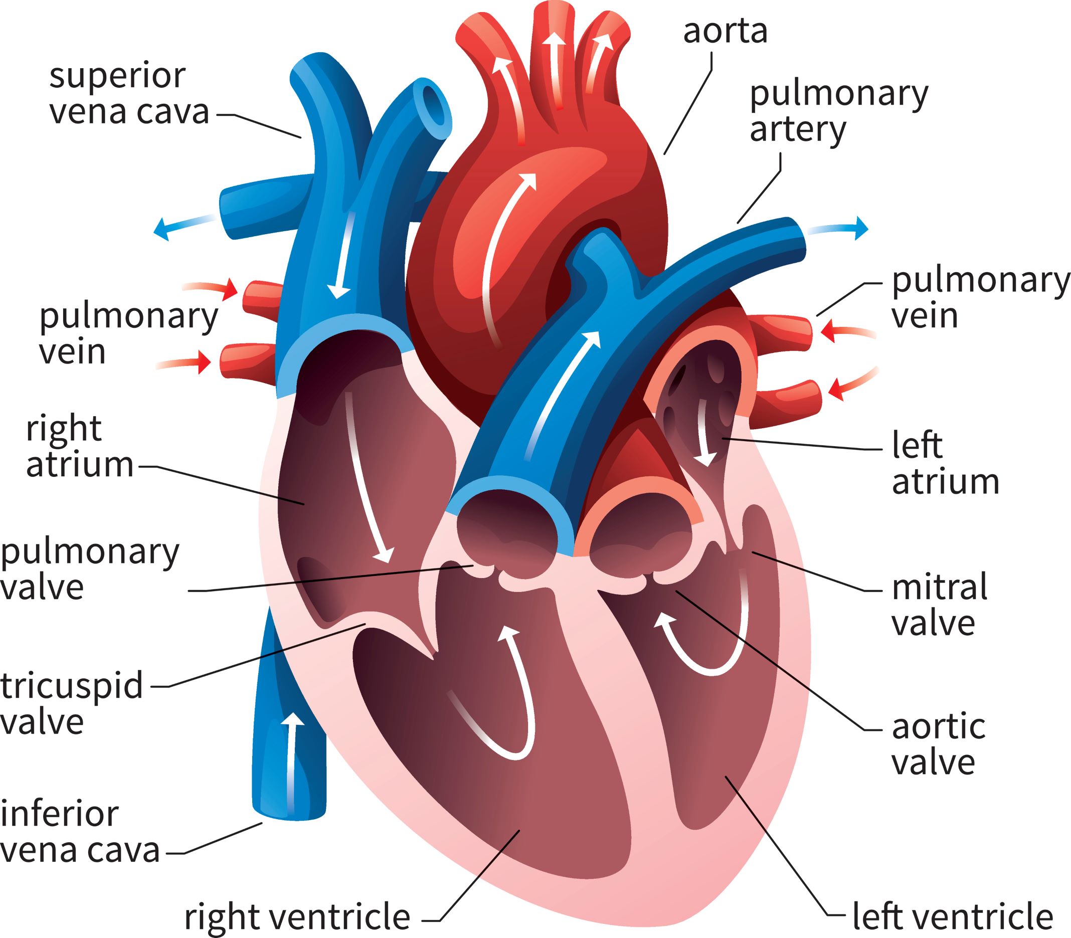
Introduction to Anatomy of the Heart This course is designed to give you a comprehensive introduction to the anatomy of the heart It is an interactive, lecture based course covering the underlying concepts and principles related to human gross anatomy of the heart and related structuresShare on A diagram of the heart's valves Image credit OpenStax College, Anatomy & Physiology, 13 The heart has four valves to ensure that blood only flows in one directionEvery day, the heart pumps about 2,000 gallons (7,600 liters) of blood, beating about 100,000 times Heart Anatomy Glossary aorta the biggest and longest artery (a blood vessel carrying blood away from the heart) in the body


Human Heart Diagram And Anatomy Of The Heart Studypk Human Heart Anatomy Anatomy And Physiology Heart Diagram
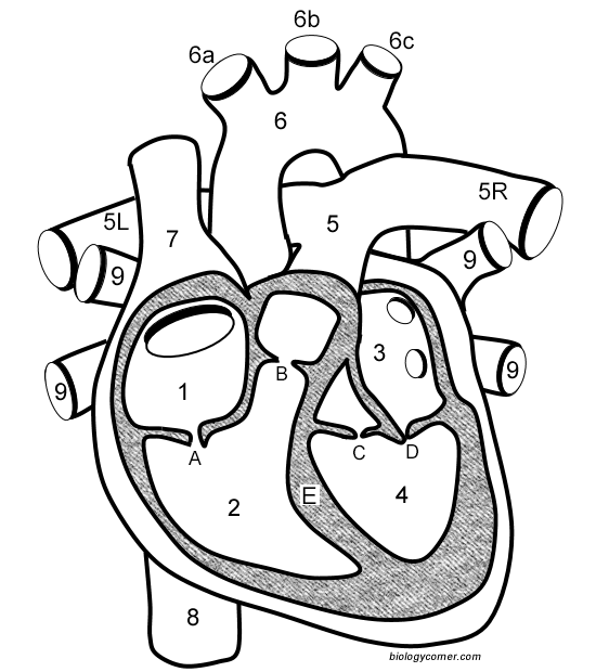


Learn The Anatomy Of The Heart
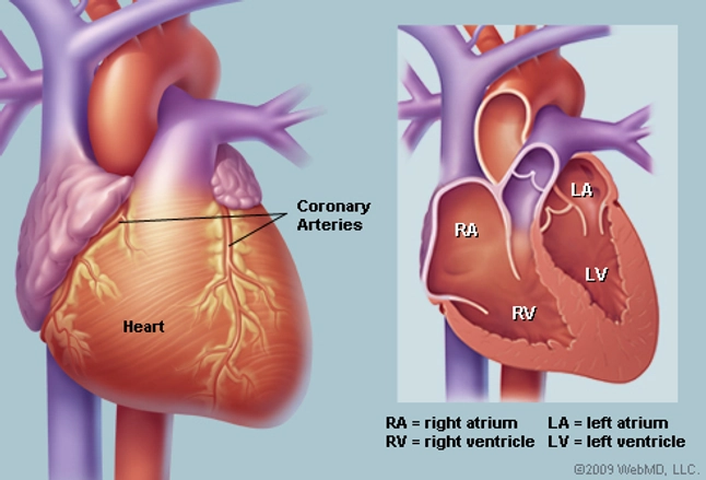
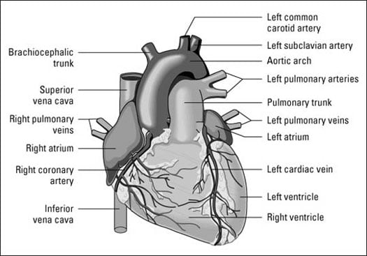
Heart Diagram Diagram of a heart Human Heart Human Heart Anatomy The human heart consists of the following parts aorta, left atrium, right atrium, left ventricle, right ventricle, veins, arteries and others Heart diagram with labelsIt consists of the heart, which is a muscular pumping device, and a closed system of vessels called arteries, veins, and capillaries As the name implies, blood contained in the circulatory system is pumped by the heart around a closed circle or circuit of vessels as it passes again and again through the various circulations of the body (on pHuman body, the physical substance of the human organism Characteristic of the vertebrate form, the human body has an internal skeleton with a backbone, and, as with the mammalian form, it has hair and mammary glands Learn more about the composition, form, and physical adaptations of the human body
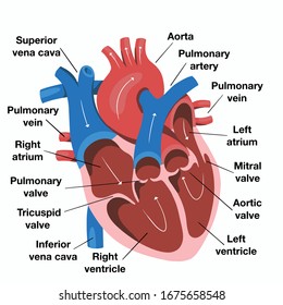


Heart Anatomy Images Stock Photos Vectors Shutterstock
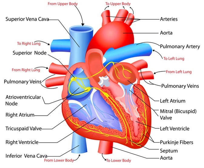


Structure And Function Of The Heart
The heart is the organ that helps supply blood and oxygen to all parts of the body Heart anatomy focuses on the structure and function of the heartMost heart attacks happen when a piece of this plaque breaks off A blood clot forms around the brokenoff plaque, and it blocks the artery Swipe to advance 3 / 10 SymptomsEvery day, the heart pumps about 2,000 gallons (7,600 liters) of blood, beating about 100,000 times Heart Anatomy Glossary aorta the biggest and longest artery (a blood vessel carrying blood away from the heart) in the body


Heart Anatomy Part Of The Human Heart Royalty Free Cliparts Vectors And Stock Illustration Image


Pictures Of Human Heart Anatomy Anatomy Of The Human Heart 4k Ultra Hd Wallpaper Human Heart Anatomy Human Anatomy And Physiology Heart Anatomy
8 anatomical planes and directions Do you know the language of anatomy?Label Heart Anatomy Diagram Printout Shark Heart and Circulatory System Shark Anatomy Heart Anatomy Glossary Printout Label Lungs Diagram Printout Label the Spine Printout Today's featured page Growing and Changing, A Printable FillintheBlanks BookIn order to understand how that happens, it is necessary to understand the anatomy and physiology of the heart Location of the Heart The human heart is located within the thoracic cavity, medially between the lungs in the space known as the mediastinum Figure 1 shows the position of the heart within the thoracic cavity


Heart Anatomy Illustration Stock Image C046 1442 Science Photo Library
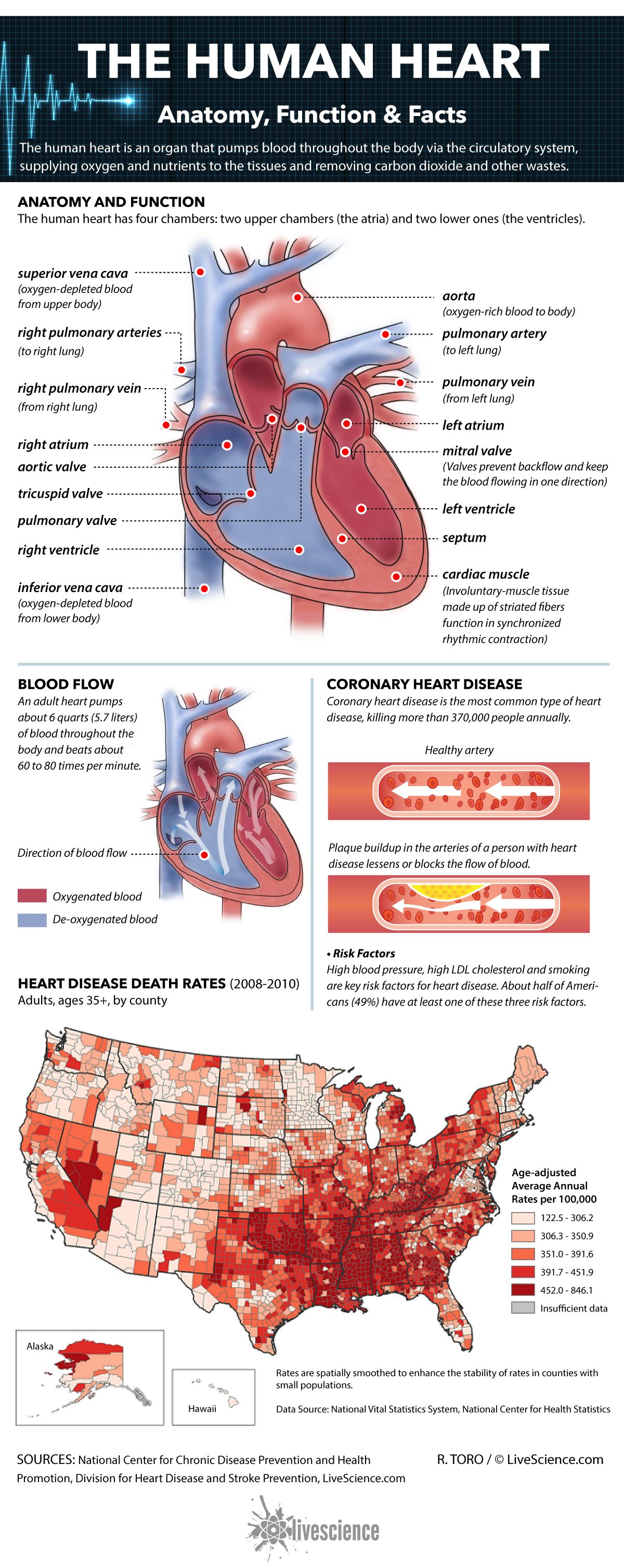


Human Heart Anatomy Function Facts Live Science
6 the heart name the parts of the human heart 7 the muscles Can you identify the muscles of the body?Start studying Heart Anatomy (Inside) Learn vocabulary, terms, and more with flashcards, games, and other study tools9 the spine Test your knowledge of the bones of the spine 10 the skin understand the functions of the integumentary system Anatomy Physiology Therapies


Q Tbn And9gcr Tz1njqe8ssrfjfyqlj9jjcr Ufmnfukbilaehxpdwujznr2 Usqp Cau
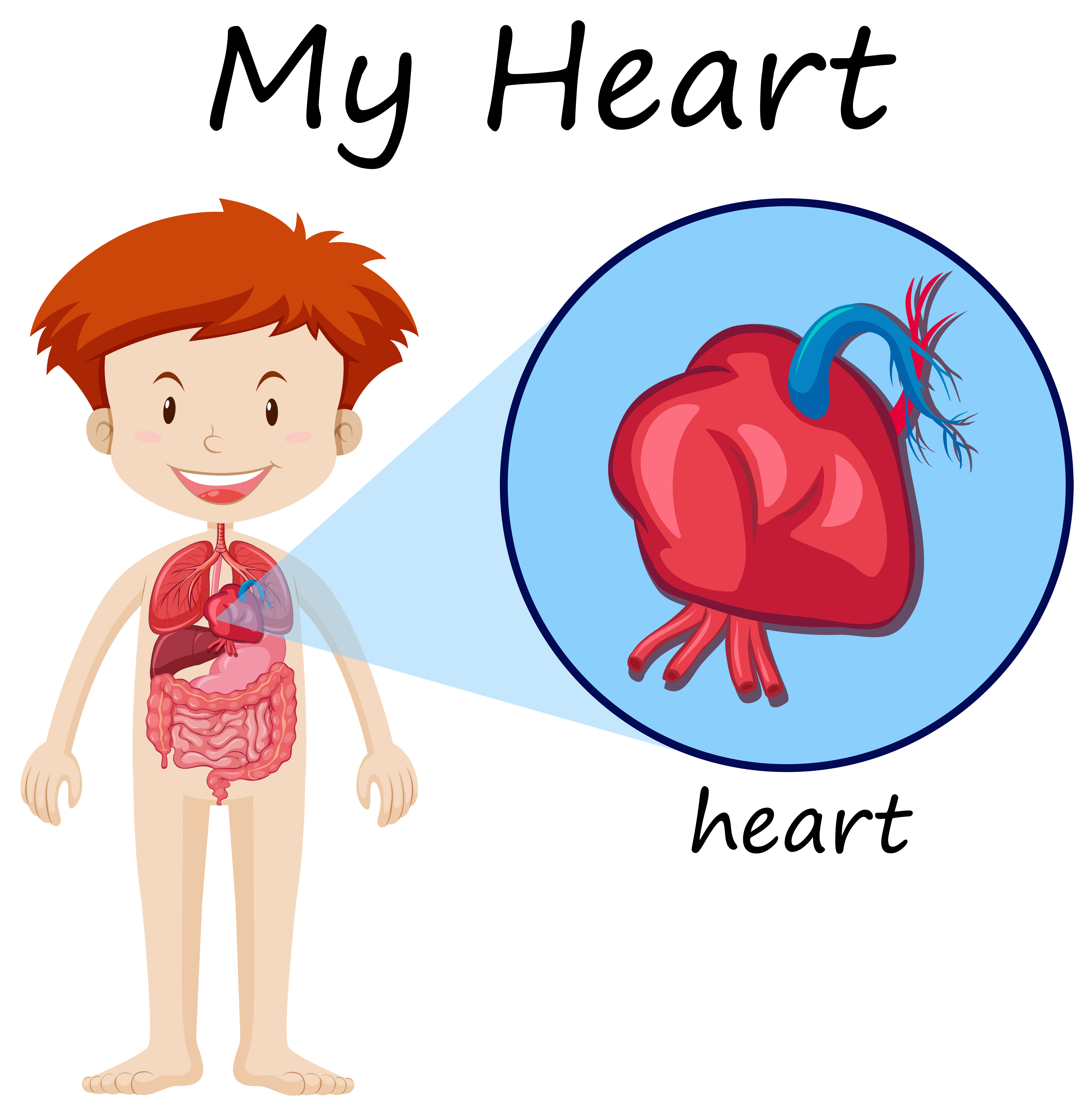


Human Anatomy Diagram With Boy And Heart Download Free Vectors Clipart Graphics Vector Art
It consists of the heart, which is a muscular pumping device, and a closed system of vessels called arteries, veins, and capillaries As the name implies, blood contained in the circulatory system is pumped by the heart around a closed circle or circuit of vessels as it passes again and again through the various circulations of the body (on pLabel Heart Anatomy Diagram Printout Shark Heart and Circulatory System Shark Anatomy Heart Anatomy Glossary Printout Label Lungs Diagram Printout Label the Spine Printout Today's featured page Growing and Changing, A Printable FillintheBlanks BookHeart Diagram Diagram of a heart Human Heart Human Heart Anatomy The human heart consists of the following parts aorta, left atrium, right atrium, left ventricle, right ventricle, veins, arteries and others Heart diagram with labels


Normal Anatomy Of The Human Heart


Human Heart Anatomy Vector Illustration Stock Vector Crushpixel
The below heart diagram with labels can be used for kids education, science classes, etc Diagram Chart Human body anatomy diagrams and charts with labels This diagram depicts Heart Diagram Human anatomy diagrams show internal organs, cells, systems, conditions, symptoms and sickness information and/or tips for healthy living Heart DiagramThe heart has four chambers, and most diagrams will show the heart as it is viewed from the ventral side This means that as you look at the heart, the left side refers to the "patient's" left side and not your left side **For each of the numbers described below, LABEL on the heart diagram**Human's heart is an amazing organ It continuously pumps oxygen and nutrientrich blood throughout your body to sustain life This fistsized powerhouse beats (expands and contracts) 100,000 times per day, pumping five or six quarts of blood each minute, or about 2,000 gallons per day
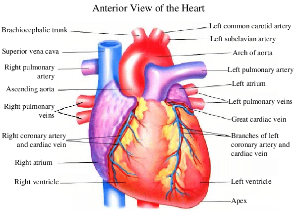


1 Heart Anatomy From The Anterior View Left And Interior View Download Scientific Diagram
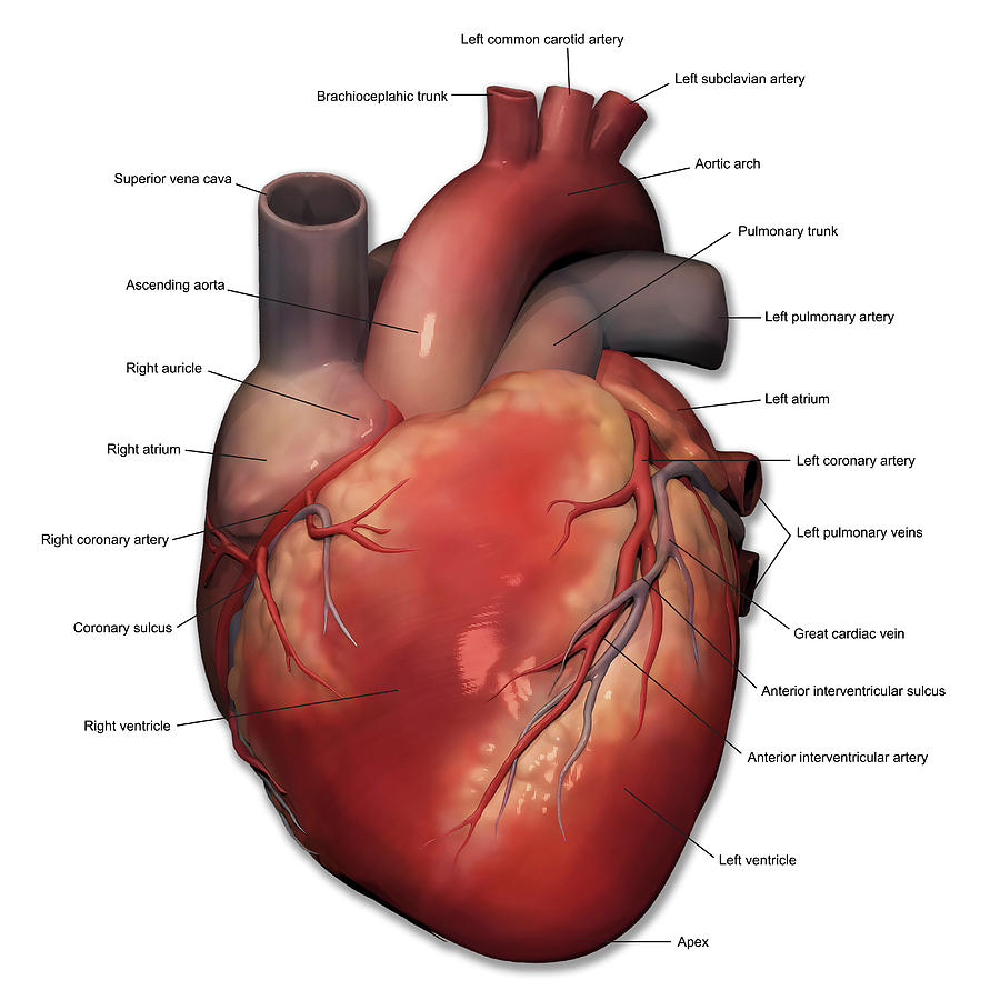


Anterior View Of Human Heart Anatomy Photograph By Alayna Guza
In this interactive, you can label parts of the human heart Drag and drop the text labels onto the boxes next to the heart diagram If you want to redo an answer, click on the box and the answer will go back to the top so you can move it to another boxHeart anatomy on different vertebrates The basic vertebrate cardiovascular system includes a heart that contracts to propel blood out to the body through arteries, and a series of blood vessels The blood enters the heart through the upper chamber(s), the atrium (or atria) Passing through a valve, blood enters the lower chamber(s), theAnatomy and Function of the Coronary Arteries Facebook Twitter Linkedin Print This can lead to a heart attack and possibly death Atherosclerosis (a buildup of plaque in the inner lining of an artery causing it to narrow or become blocked) is the most common cause of heart disease


Heart Detail Picture Image On Medicinenet Com
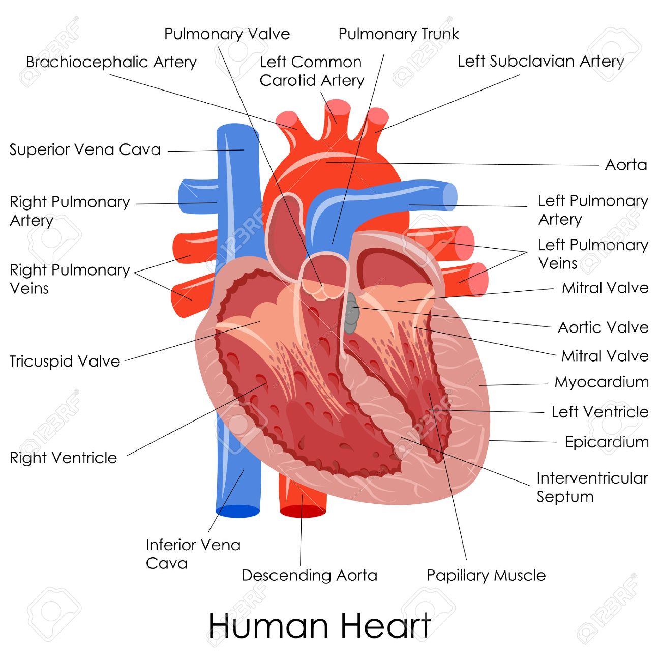


Vector Illustration Of Diagram Of Human Heart Anatomy Stock Photo Picture And Royalty Free Image Image
Anatomy of the human heart and coronaries how to view anatomical structures This tool provides access to an MDCT atlas in the 4 usual planes, allowing the user to interactively discover the heart anatomy The images are labeled, providing an important medical and anatomical tool The quiz mode makes it possible to evaluate the user's progressAnatomy of the human heart and coronaries how to view anatomical structures This tool provides access to an MDCT atlas in the 4 usual planes, allowing the user to interactively discover the heart anatomy The images are labeled, providing an important medical and anatomical tool The quiz mode makes it possible to evaluate the user's progressHeart Anatomy Your heart is located between your lungs in the middle of your chest, behind and slightly to the left of your breastbone (sternum) A doublelayered membrane called the pericardium surrounds your heart like a sac The outer layer of the pericardium surrounds the roots of your heart's major blood vessels and is attached by


Pin On Homeschool Idea S C C Ch A Help
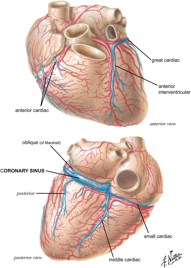


Anatomy Of The Human Heart Springerlink
Whatever you need to learn, use our intuitive atlas to effortlessly understand the topics you've always struggled with Start learning anatomy in less than 60 secondsWebMD's Heart Anatomy Page provides a detailed image of the heart and provides information on heart conditions, tests, and treatmentsHeart Anatomy An online interactive study guide to tutorials and quizzes on the anatomy and physiology of the heart, using interactive animations and diagrams Looking for free labeling diagrams?


Human Heart Diagram And Anatomy Of The Heart Anatomy And Physiology Heart Diagram Human Heart Anatomy
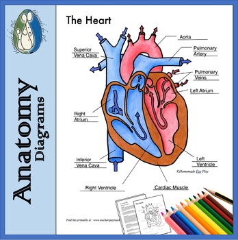


Heart Diagrams For Labeling And Coloring With Reference Chart And Summary
This MRI heart cross sectional anatomy tool is absolutely free to use Use the mouse scroll wheel to move the images up and down alternatively use the tiny arrows (>>) on both side of the image to move the images>>) on both side of the image to move the imagesFrom human body organ diagrams to skull bones and chambers of the heart;The heart wall itself can be divided into three distinct layers the endocardium, myocardium, and epicardium In this article, we shall look at the anatomy and clinical relevance of these layers Endocardium The innermost layer of the cardiac wall is known as the endocardium It lines the cavities and valves of the heart


Human Heart Anatomy Pre Designed Photoshop Graphics Creative Market


3
Multiple choice questions on the Anatomy of the heart include few questions on the Anatomy of the heart at the high school level It is useful for premedical entrance examination as wellThe human heart is roughly heartshaped structure and rests obliquely in the thoracic cavity (Fig 73) The anterior surface of the human heart faces the sternum, the posterior surface—the base of the cone faces the vertebral column and the inferior or diaphragmatic surface rests on the diaphragmA heart diagram is a popular design used by different people for various uses It can be used by a teacher or student for academic purpose, by a friend or relative for mutually sending and exchanging cards or for baby toys or printing on dresses etc


Human Heart Anatomy Blood Flow Stock Illustration Download Image Now Istock


The Human Heart Anatomy Passage Of Blood Teachpe Com
The heart separates the pulmonary and systemic circulations, which ensures the flow of oxygenated blood to tissues Anatomy of the Heart The cardiovascular system can be compared to a muscular pump equipped with oneway valves and a system of large and small plumbing tubes within which the blood travelsLearn anatomy human heart diagrams with free interactive flashcards Choose from 500 different sets of anatomy human heart diagrams flashcards on QuizletThe heart wall itself can be divided into three distinct layers the endocardium, myocardium, and epicardium In this article, we shall look at the anatomy and clinical relevance of these layers Endocardium The innermost layer of the cardiac wall is known as the endocardium It lines the cavities and valves of the heart


Heart Anatomy Fill In The Blank Diagram Quizlet


Heart Anatomy Wallpapers Wallpaper Cave
The heart is made of three layers of tissue Endocardium, the thin inner lining of the heart chambers that also forms the surface of the valves Myocardium, the thick middle layer of muscle that allows your heart chambers to contract and relax to pump blood to your body Pericardium, the sac that surrounds your heart Made of thin layers of tissue, it holds the heart in place and protects it9 the spine Test your knowledge of the bones of the spine 10 the skin understand the functions of the integumentary system Anatomy Physiology TherapiesThe heart is a mostly hollow, muscular organ composed of cardiac muscles and connective tissue that acts as a pump to distribute blood throughout the body's tissues


Human Heart Muscle Structure Anatomy Diagram Infographic Chart Diagram With All Parts Outside View Right Left Atrium Canstock


Heart Diagram Anatomy Diagram Blue Food Chicken Png Pngwing
Ummmmmmm it's pretty self explanatory you label the heart Just remember one thing you're looking at the heart like it's in someone else so right and left are switched aroundFind human heart diagram stock images in HD and millions of other royaltyfree stock photos, illustrations and vectors in the collection Thousands of new, highquality pictures added every day6 the heart name the parts of the human heart 7 the muscles Can you identify the muscles of the body?


Sketch Of Human Heart Anatomy With Hand Written Labels Stock Illustration Download Image Now Istock


Human Heart Anatomy System Human Body Anatomy Diagram And Chart Images
8 anatomical planes and directions Do you know the language of anatomy?
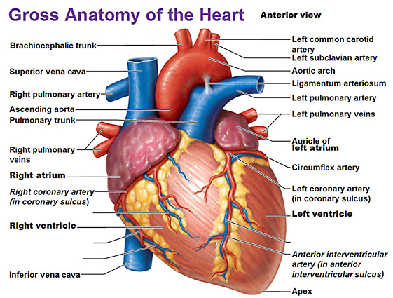


Heart Anatomy


Chapter 27 Heart Anatomy Bio 140 Human Biology I Textbook Libguides At Hostos Community College Library


28 Collection Of Drawing Of A Human Heart And Its Parts Simple Heart Anatomy Diagram Png Image Transparent Png Free Download On Seekpng


Anatomical Heart Diagram Black Text Art Print By Dianeleonardart Redbubble


Heart Anatomy Anatomy And Physiology


Know Your Heart Anatomy 101 Cardiac Heart Health Health Topics Hackensack Meridian Health
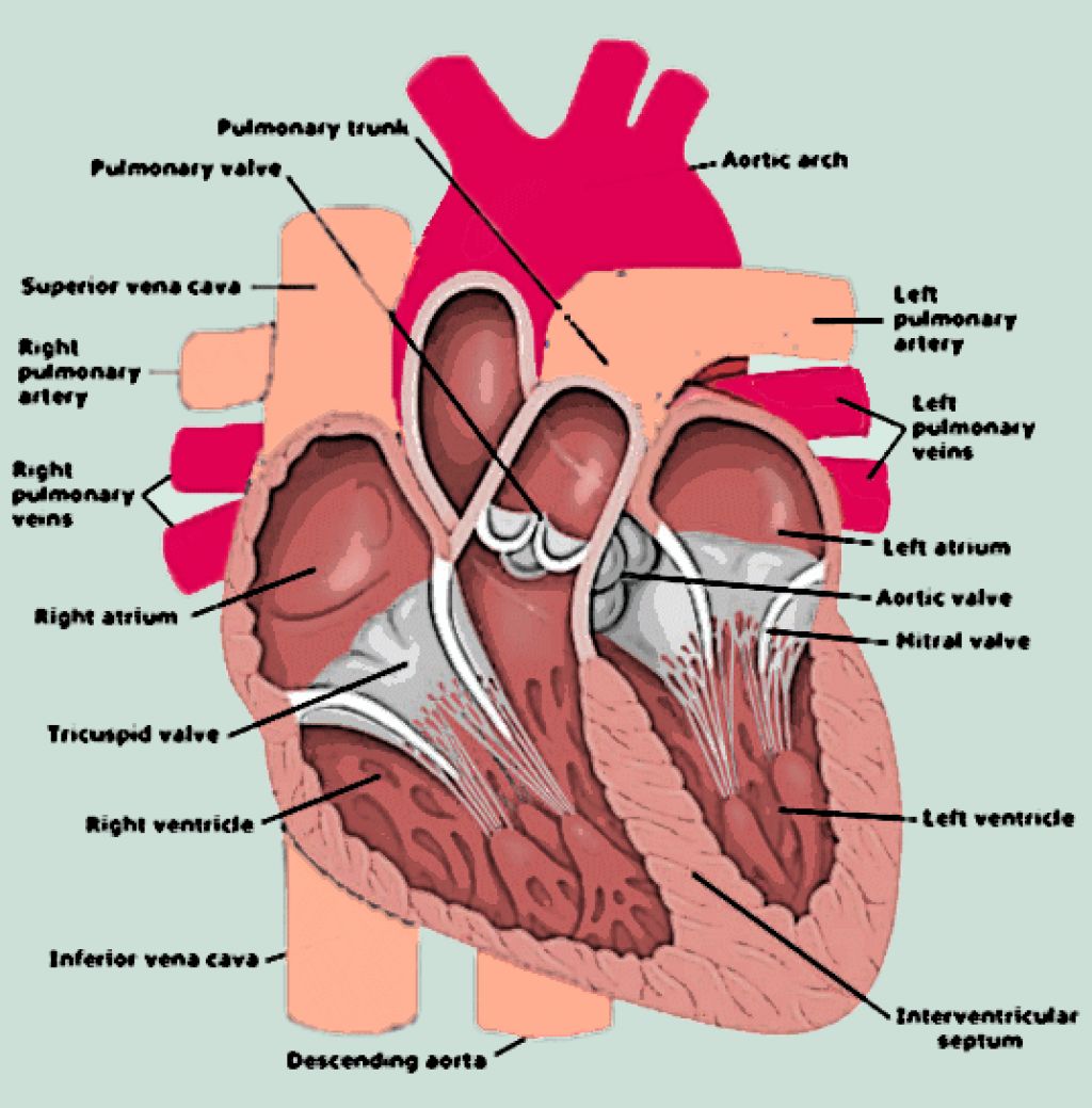


Anatomy Of Human Heart Diagram Free Image


Human Heart Anatomy Diagram Function Chambers Location In Body


Basic Anatomy Of The Human Heart Cardiology Associates Of Michigan Michigan S Best Heart Doctors


Figuring Out Cardiac Anatomy Your Heart Dummies


Normal Heart Anatomy And Blood Flow Pediatric Heart Specialists
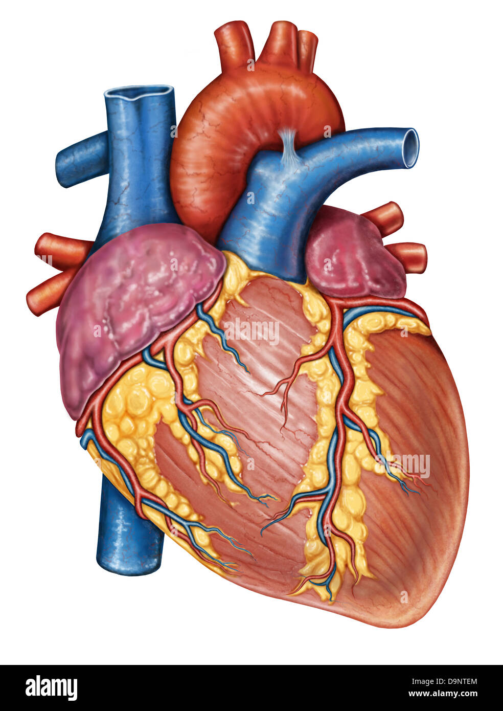


Gross Anatomy Of The Human Heart Stock Photo Alamy
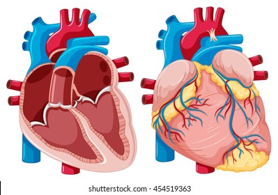


Heart Anatomy Images Stock Photos Vectors Shutterstock


Anatomy Of A Human Heart


Heart Structure Function Facts Britannica
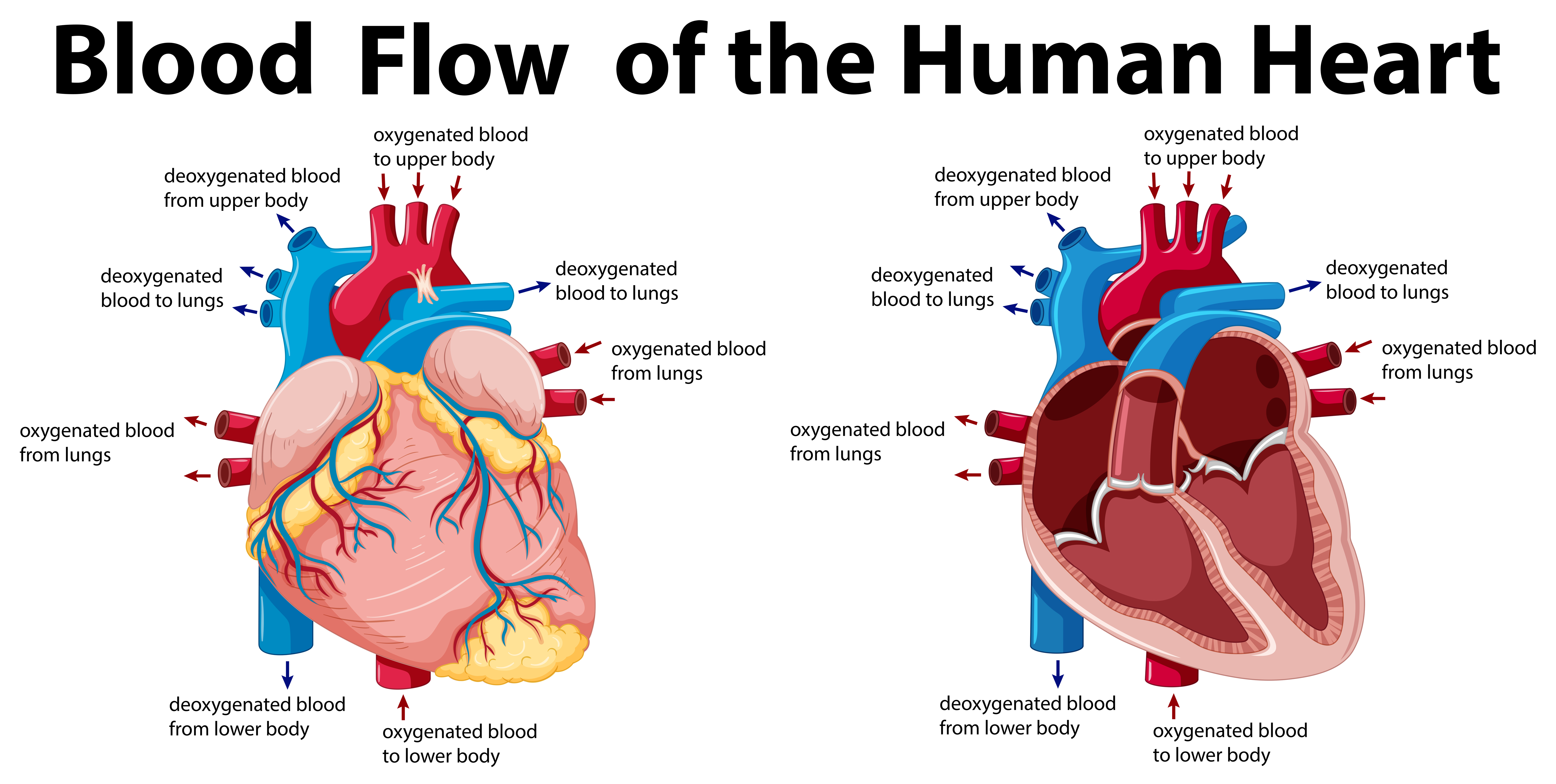


Blood Flow Of The Human Heart Download Free Vectors Clipart Graphics Vector Art
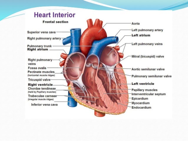


Human Heart Anatomy And Physiology Part 1
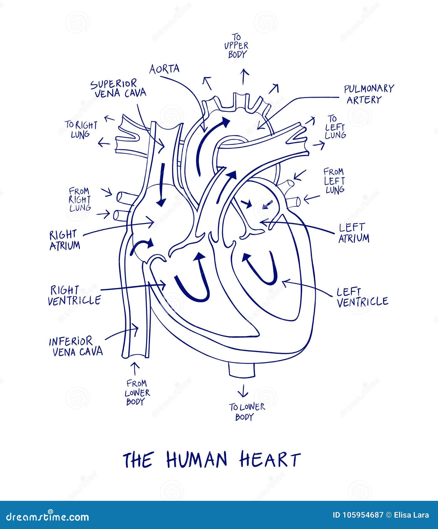


Sketch Of Human Heart Anatomy On Blue Line On A White Background Stock Vector Illustration Of Cardiac Heartbeat


Cardiac Anatomy And Electrophysiology Thoracic Key
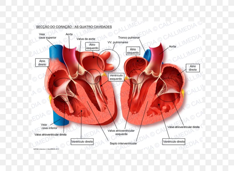


Human Heart Anatomy Blood Vessel Pulmonary Vein Png 600x600px Watercolor Cartoon Flower Frame Heart Download Free
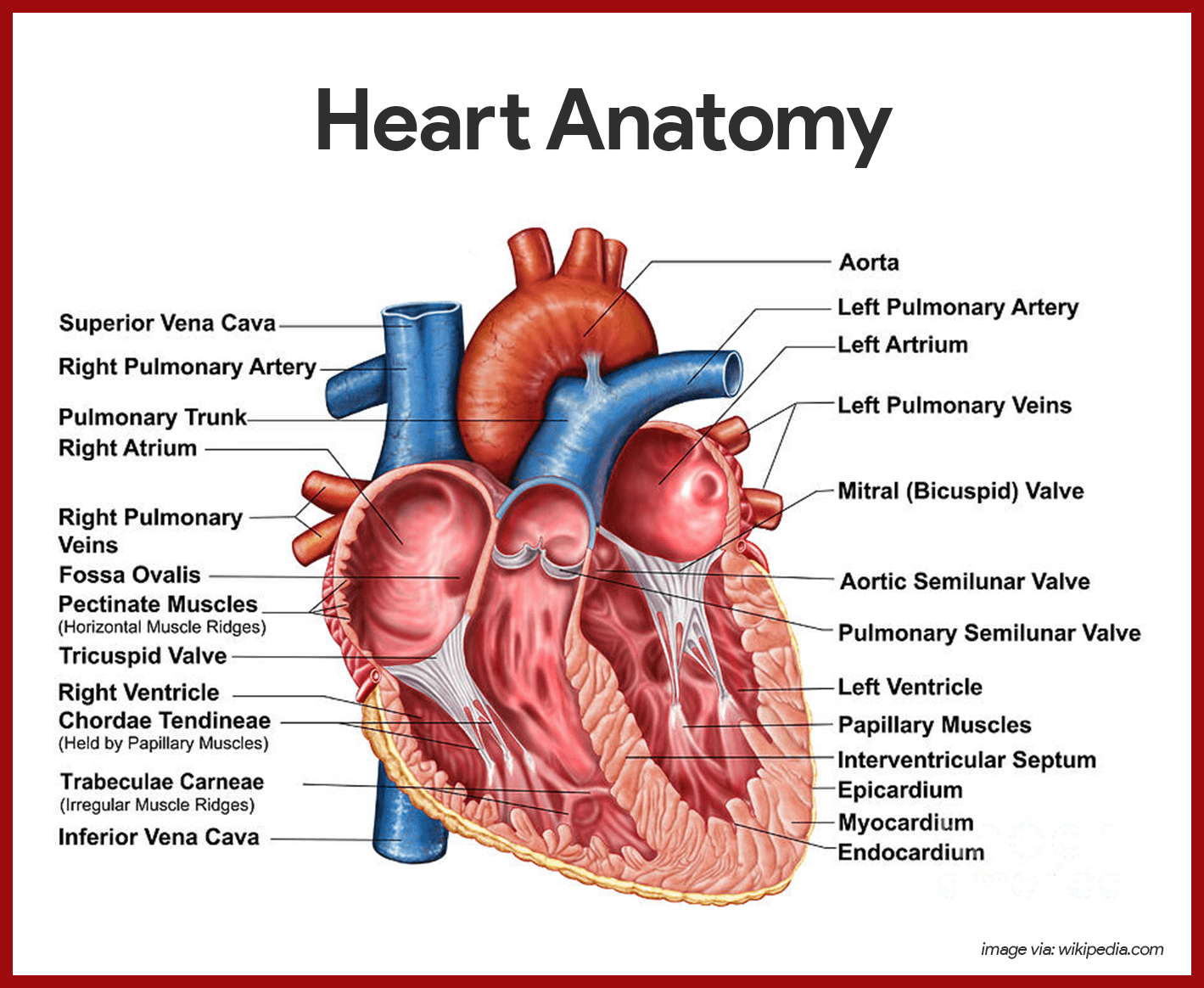


Cardiovascular System Anatomy And Physiology Study Guide For Nurses


Label Heart Anatomy Diagram Printout Enchantedlearning Com


Anatomical Diagrams Of Heart Heart Failure Online
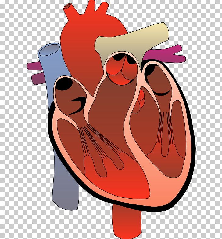


Heart Anatomy Diagram Circulatory System Png Clipart Anatomy Art Beak Bird Cartoon Free Png Download
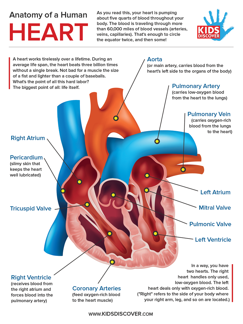


Infographic Anatomy Of The Human Heart Kids Discover


How The Heart Works Diagram Anatomy Blood Flow


Anatomy Of A Human Heart Anatomy Drawing Diagram


Anatomy Of The Heart Emt Training Station


Human Heart Muscle Structure Anatomy Diagram Art Print Barewalls Posters Prints Bwc


Physiology Tutorial The Human Heart
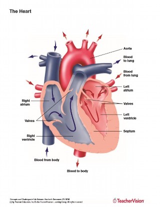


Anatomy Of The Human Heart Printable 6th 12th Grade Teachervision


Q Tbn And9gctjrns7oisdp0o1qlhr2hfhxrxtvv 7rxgfnkpv Oyspdj1rkoi Usqp Cau


The Normal Heart


Coronary Circulation Wikipedia


Heart Anatomy Mydr Com Au
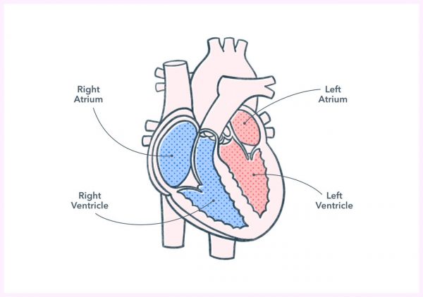


Anatomy And Physiology Of The Human Heart Pocket Prep
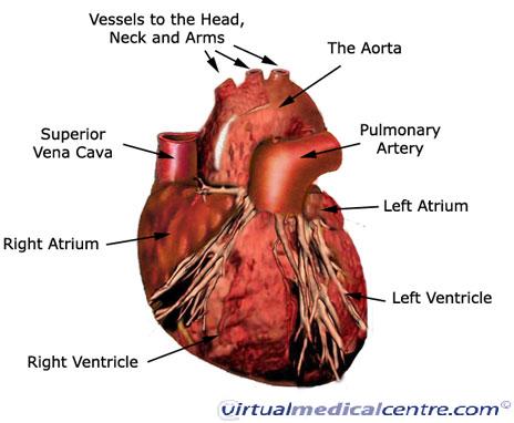


Cardiovascular System Heart Anatomy Healthengine Blog


Anatomy Of A Human Heart Bibloteka
:background_color(FFFFFF):format(jpeg)/images/library/10912/labeled_heart_diagram.png)


Diagrams Quizzes And Worksheets Of The Heart Kenhub
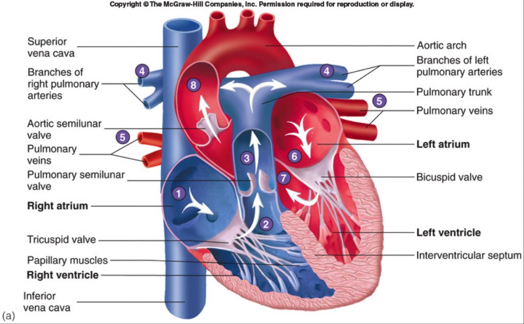


Human Heart Gross Structure And Anatomy Online Biology Notes


Human Heart Anatomy Schematic Diagram Vector Illustration Royalty Free Cliparts Vectors And Stock Illustration Image
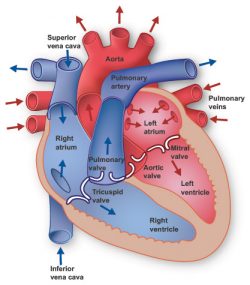


Heart Information Center Heart Anatomy Texas Heart Institute
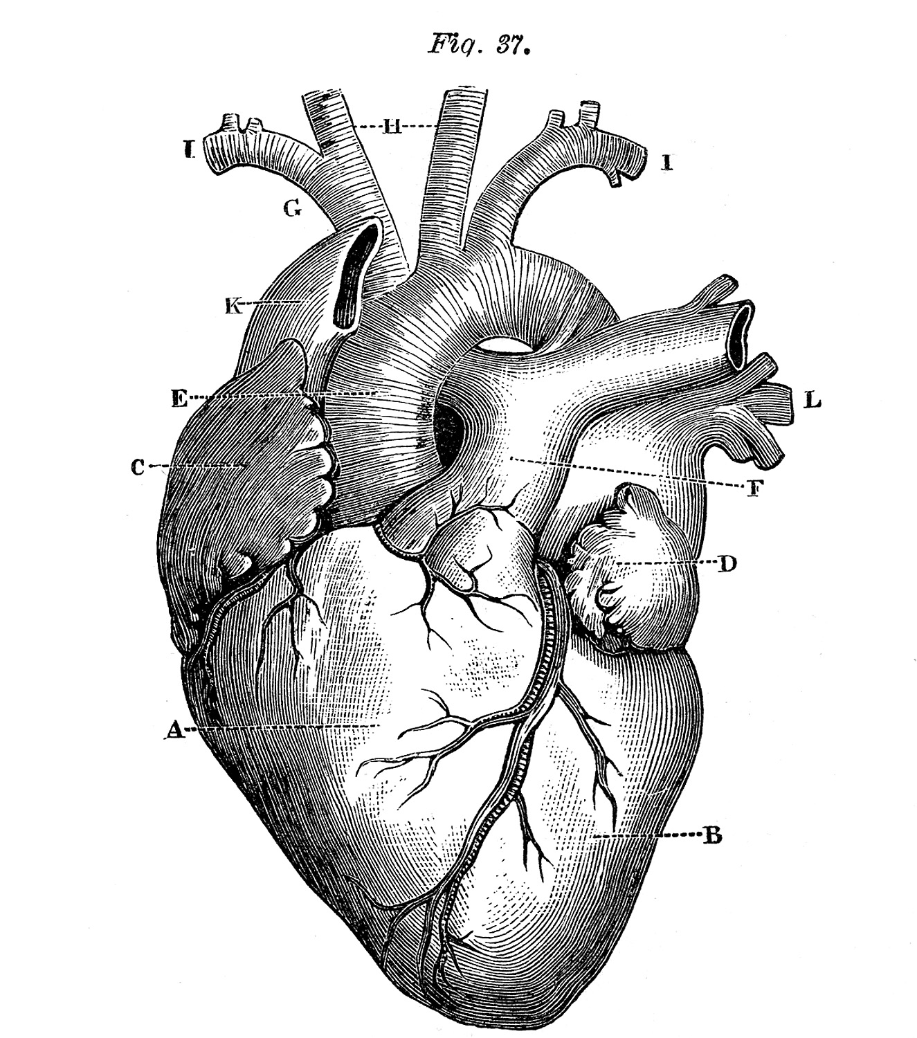


6 Anatomical Heart Pictures The Graphics Fairy


Heart Anatomy Lab Quiz Interior View Diagram Quizlet


1 Shows The Heart Anatomy From The Anterior And Interior Views The Download Scientific Diagram


Anatomy Of The Heart Powerpoint Template Free Download Powerpoint University Youtube
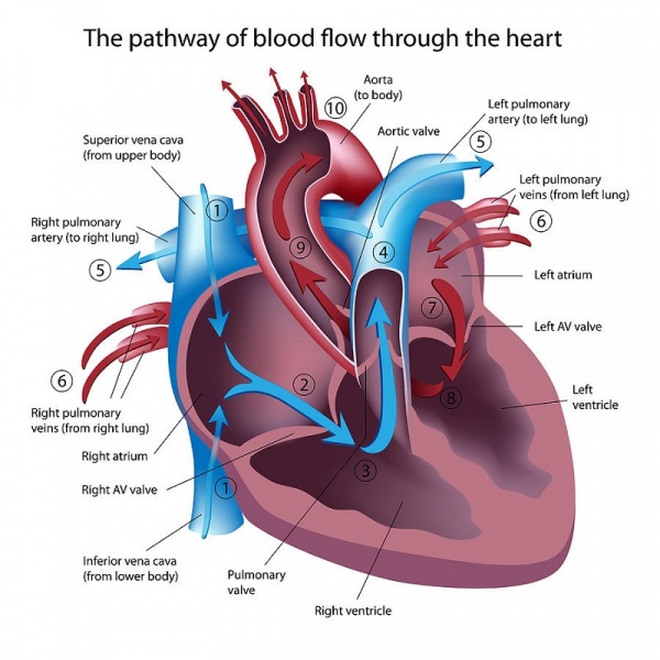


Anatomy Of The Human Heart Physiopedia
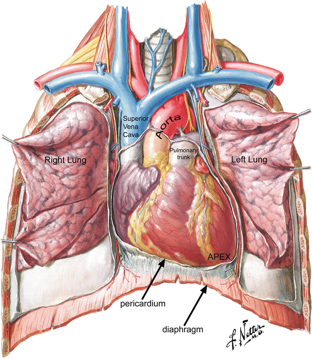


Anatomy Of The Human Heart Springerlink


Heart Anatomy Yourheartvalve


Anatomy Of The Human Heart Internal Structures


Anatomy Of The Human Heart
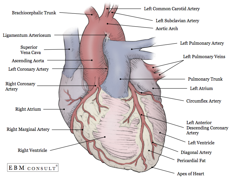


Anatomy Heart External


Openstax Anatomy And Physiology Ch19 The Cardiovascular System The Heart Top Hat Cardiac Anatomy Heart Diagram Heart Anatomy
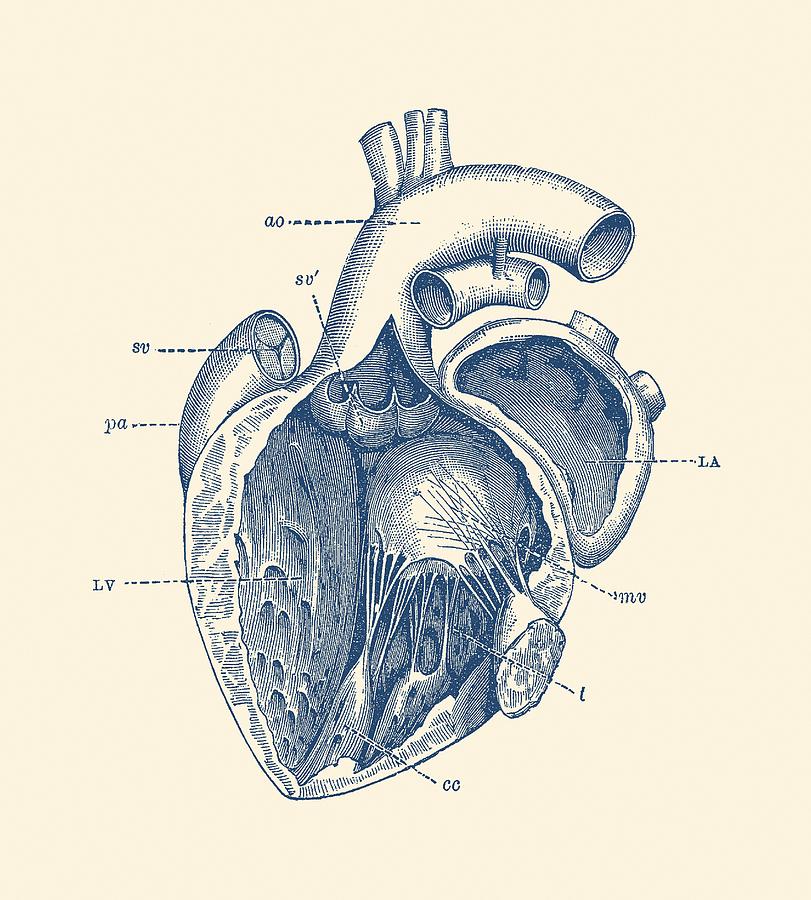


Internal Human Heart Diagram Anatomy Poster Drawing By War Is Hell Store


Q Tbn And9gcqpvnsfyxwvjlbxhq9nnfcnbhwnxd 1blnfmtetnug2ln22nuku Usqp Cau
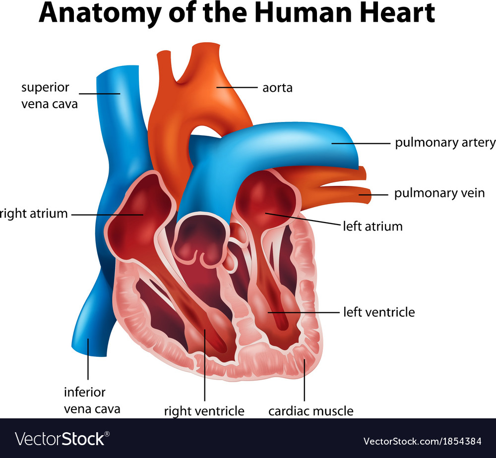


Human Heart Anatomy Royalty Free Vector Image Vectorstock
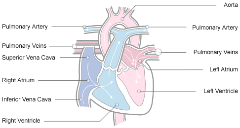


Anatomy And Physiology Of The Heart Normal Function Of The Heart Cardiology Teaching Package Practice Learning Division Of Nursing The University Of Nottingham
/heart_inner_section-577d5c673df78cb62c939314.jpg)


Atria Of The Heart Function


Human Heart Diagram Vintage Anatomy Postcard By Vaposters Redbubble


Seer Training Structure Of The Heart


Anatomy Of A Human Heart


Heart Anatomy Glossary Printout Enchantedlearning Com
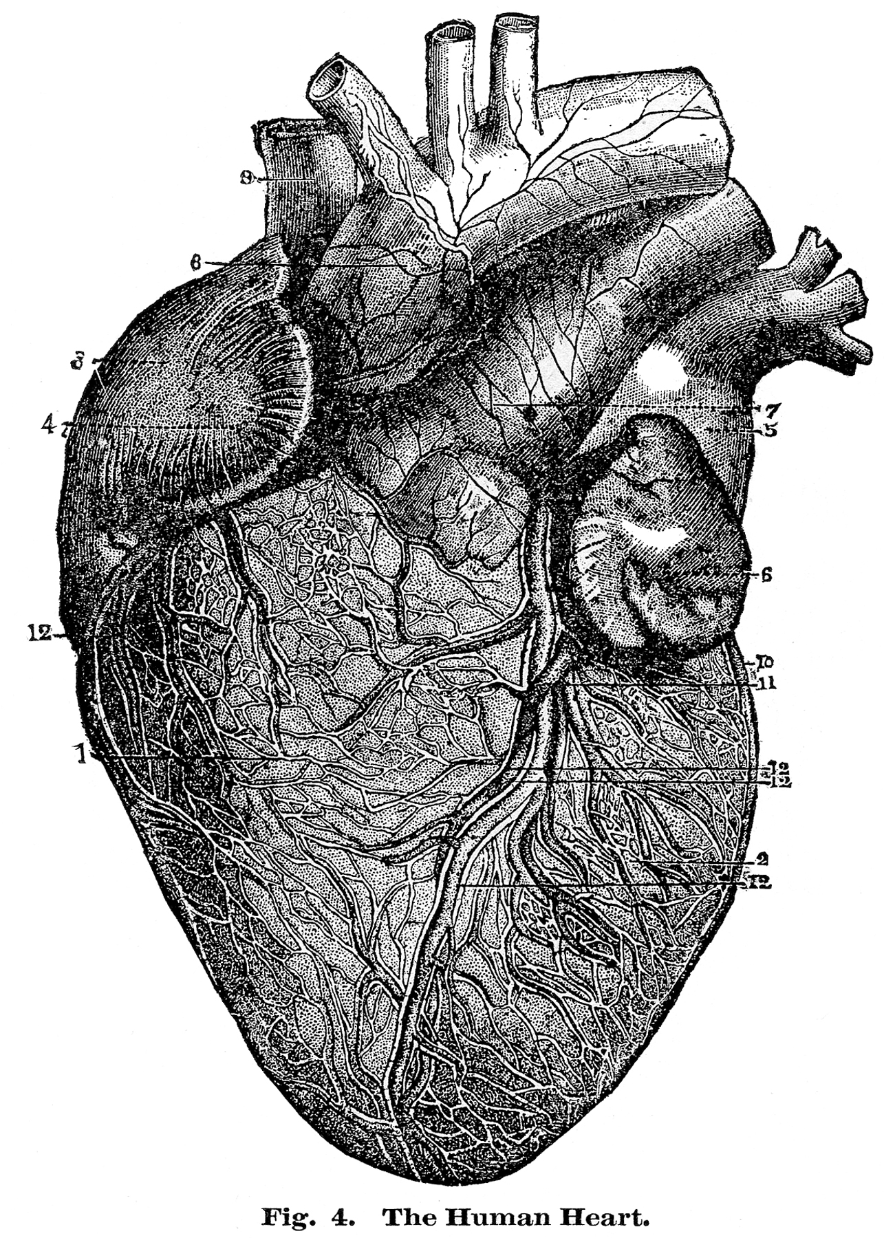


6 Anatomical Heart Pictures The Graphics Fairy
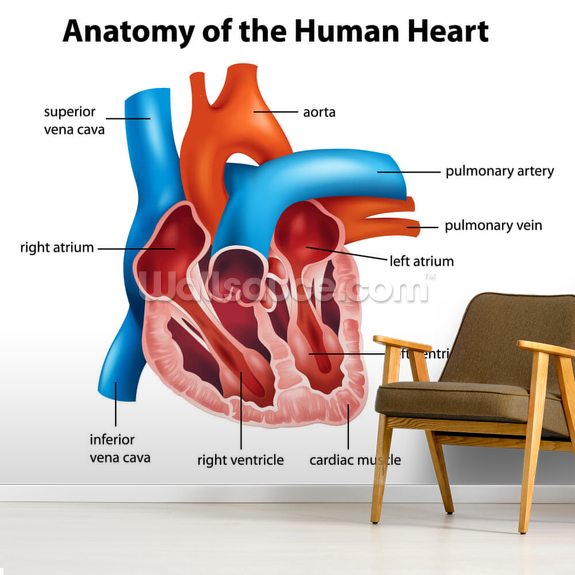


Human Heart Anatomy Wallpaper Mural Wallsauce Us
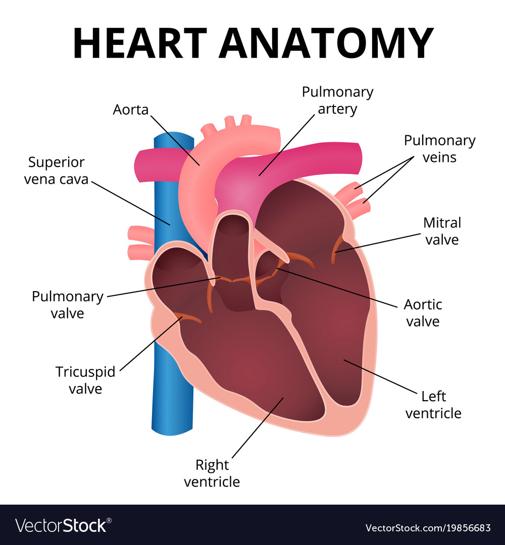


Diagram Of Heart Anatomy Anatomy Drawing Diagram


Atrium Heart Wikipedia


What Is The Real Cardiac Anatomy Mori 19 Clinical Anatomy Wiley Online Library
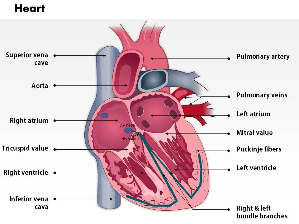


0514 Heart Anatomy Medical Images For Powerpoint Presentation Powerpoint Diagrams Ppt Sample Presentations Ppt Infographics


The Heart Anatomy How It Works And More


Anatomy Of The Heart Anatomy Of The Heart Diagram Human Body Heart Heart Human Png Pngegg


Anatomy Tutorial Attitudinally Correct Anatomy Atlas Of Human Cardiac Anatomy
:background_color(FFFFFF):format(jpeg)/images/library/11110/Heart_Thumbnail.png)


Heart Anatomy Structure Valves Coronary Vessels Kenhub


Heart Valve Anatomy Britannica


Amazon Com Emvency Mouse Pads Anatomical Of Diagram Human Heart Anatomy Body Cardiac Muscle Organ Mousepad 9 5 X 7 9 For Laptop Desktop Computers Accessories Mini Office Supplies Mouse Mats Office Products


コメント
コメントを投稿